• BoneFinder®

  • BoneFinder®

  • Bone shape analyses made easy!

    Automatic – Accurate – Adaptive
    Fast
    GET STARTED
  • Bone shape analyses made easy!

    Automatic – Accurate – Adaptive
    Fast
    GET STARTED

ABOUT

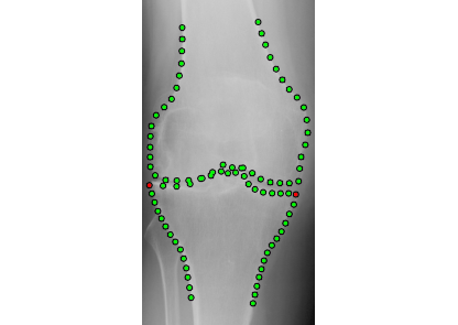
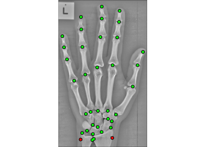
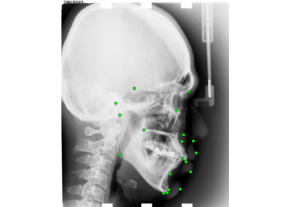
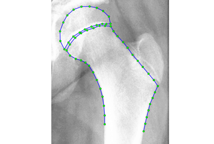
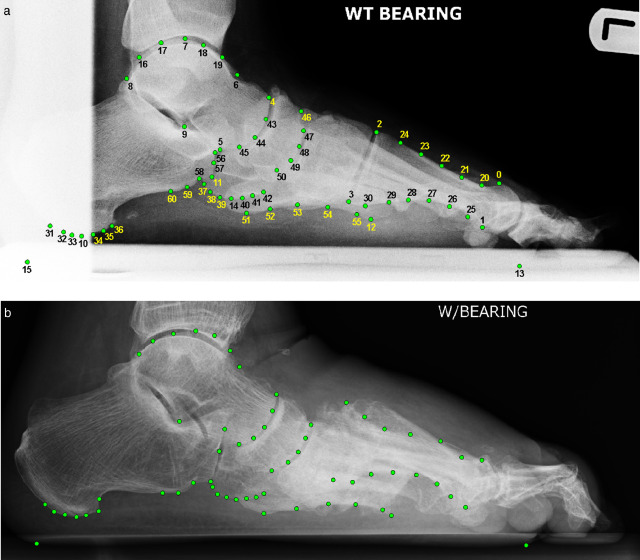
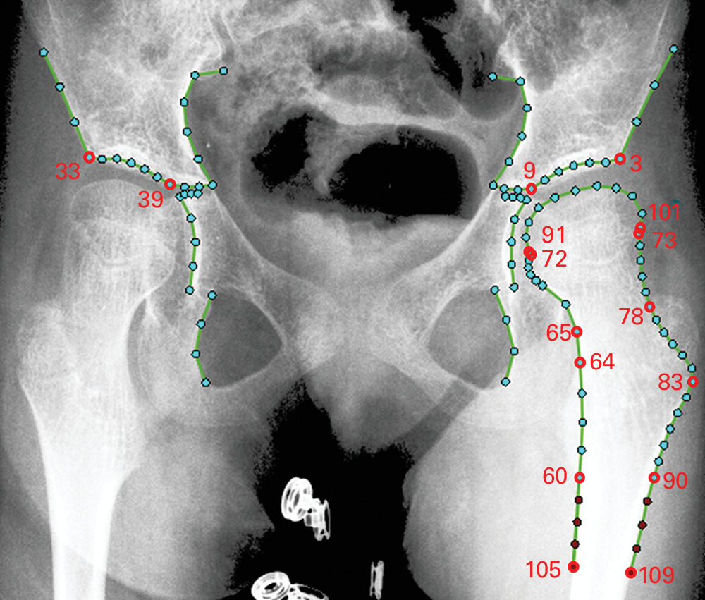
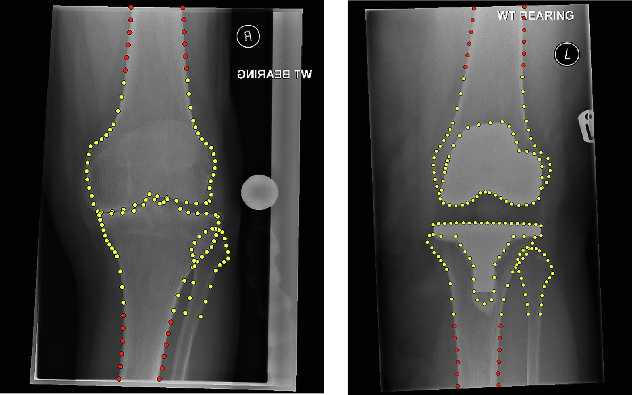
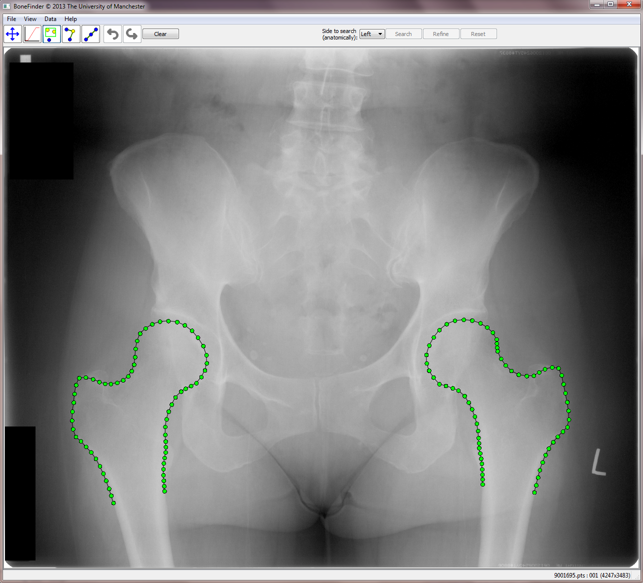
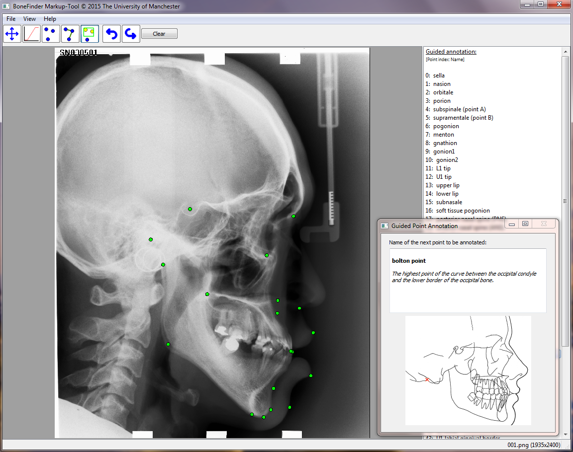
BoneFinder® is a fully automatic software tool to outline and segment skeletal structures from 2D radiographs by placing a set of points along the bone contour or at key landmark positions. It was originally designed for the segmentation of the hip joint but is now also used for other skeletal structures such as the joints of the hand or the knee joint.

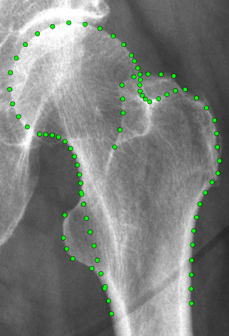
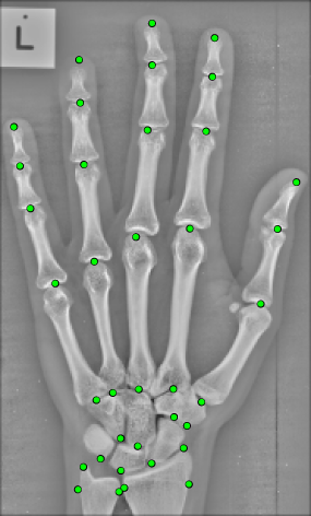
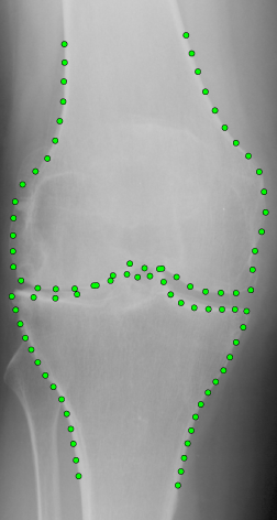
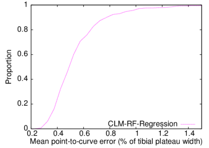
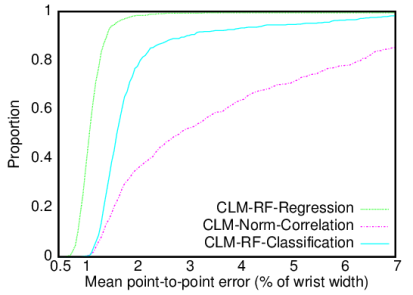
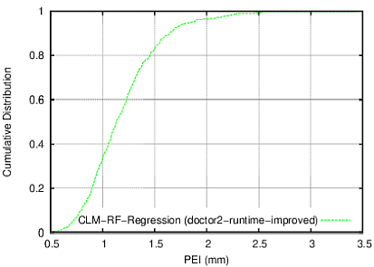
BoneFinder® firstly identifies the rough position of the bone in the image and then outlines its contour. To do so, BoneFinder® follows a machine-learning approach. This means that it has been trained on lots of examples in order to learn what to look for in an image, and it now uses this acquired knowledge to identify and segment similar bones in new unseen images. BoneFinder® has been found to lead to very robust and accurate point placements across skeletal application areas.

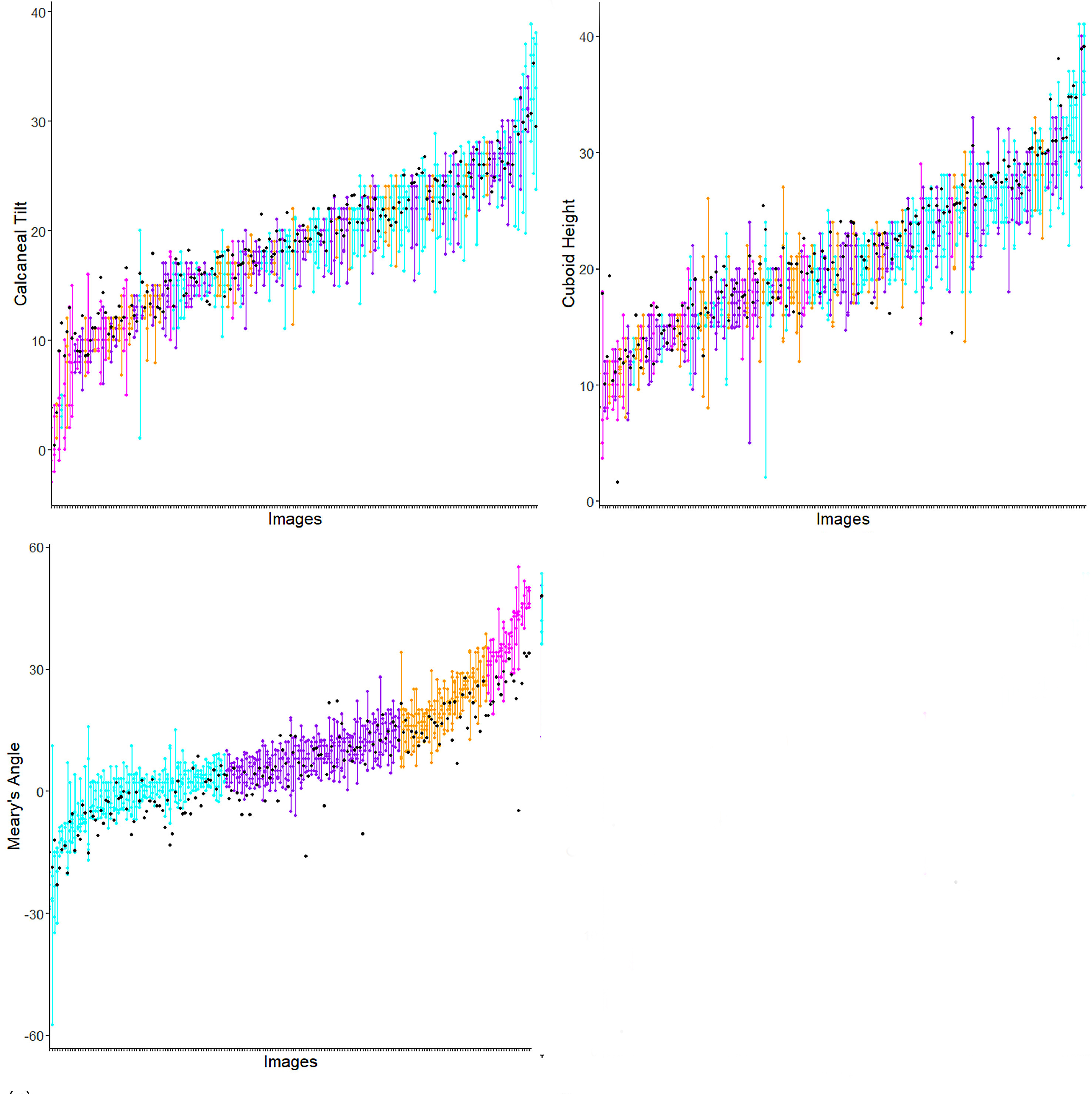
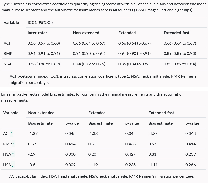
The resulting point positions can then be used for a range of shape analyses such as building statistical shape models or automatically deriving conventional geometric measurements.

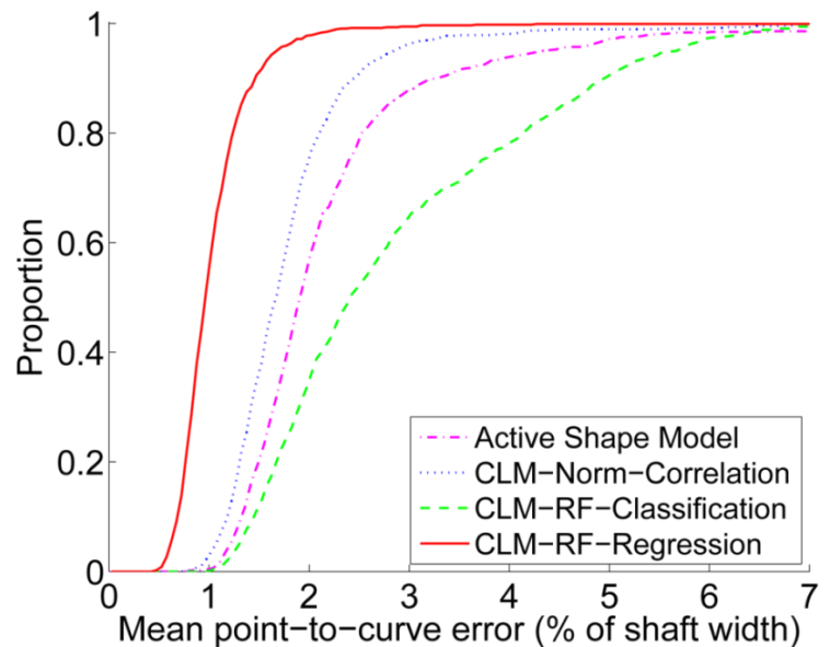
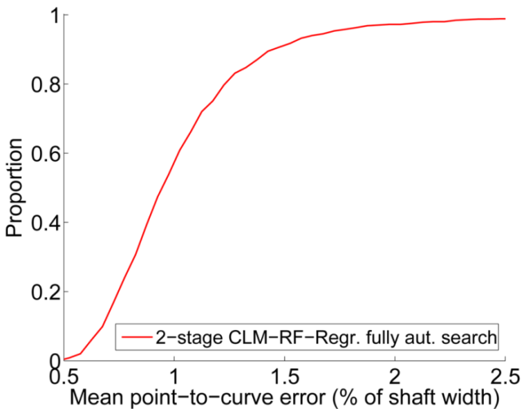
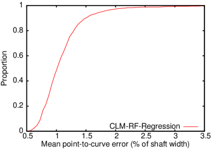
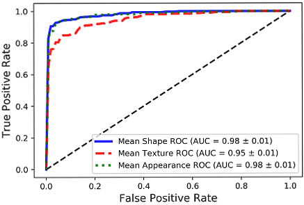
Scientific background: BoneFinder® uses a Hough Forest like approach to detect the structure of interest in the image, and then applies Random Forest Regression-Voting in the Constrained Local Model framework to locally refine all point positions. More details on these methods and relevant references can be found in our peer-reviewed journal publications of international standing in IEEE TMI and IEEE PAMI. The underlying BoneFinder®-technology has been patented: T. Cootes, C. Lindner, M. Ionita. Image processing apparatus and method for fitting a deformable shape model to an image using random forest regression voting. Patent numbers EP 2893491 (approved for GB, FR, DE), US 9928443 (approved for US).

BoneFinder® was written by Claudia Lindner, Tim Cootes and other members of the Centre for Imaging Sciences at The University of Manchester, UK. Funding for the development of BoneFinder® has been received from the Medical Research Council UK, the Engineering and Physical Sciences Research Council UK, Versus Arthritis, the Wellcome Trust and the National Institute for Health Research.

PERFORMANCE RESULTS

We tested BoneFinder® on a range of different skeletal structures using two-fold cross-validation experiments.
The results show that BoneFinder® achieves state-of-the-art performance across application areas.

We tested the BoneFinder® hip module on 839 AP pelvic radiographs (527 females, 312 males) using two-fold cross-validation experiments. The aim was to place 65 dense points along the front-view contour of the proximal femur. We report mean point-to-curve errors (where "mean" refers to averaging the errors over all points per image) as a percentage of the shaft width (based on a subset of calibrated images we estimated the latter to be 37mm).

BoneFinder® outperforms alternative matching techniques significantly when starting searching from the mean shape at true pose. It achieves a mean point-to-curve error of less than 0.9mm for 99% of all images (i.e. for 99% of images the error is less than 0.9mm).

BoneFinder® is fully automatic and highly accurate. Without any initialisation it achieves a mean point-to-curve error of less than 0.9mm for 99% of all images. This is equally good as a local search started from the mean shape at true pose (see left plot).

Examples of segmentation results of the fully automatic system (sorted by mean point-to-curve error percentiles): a) median – 0.4mm; b) 91.2% – 0.6mm, highest global search error (i.e. the global search had a success rate of 100%); c) 99.0% – 0.9mm; d) maximal overall error – 2.7mm.


Data shown on this page is copyright (c) 2013 IEEE (DOI: 10.1109/TMI.2013.2258030). Personal use of this material is permitted. However, permission to use this material for any other purposes must be obtained from the IEEE by sending a request to pubs-permissions@ieee.org.

We tested the BoneFinder® hip module on 756 AP pelvic radiographs, aiming to place 81 dense points along the contour of the proximal femur including the trochanters. We report mean point-to-curve errors (where "mean" refers to averaging the errors over all points per image) as a percentage of the shaft width (based on an average of 37mm).

The fully automatic BoneFinder® proximal femur module achieves a mean point-to-curve error of within 1.0mm for 99% of all images (i.e. for 99% of images the error is within 1.0mm).

Segmentation example showing the 95%ile of the fully automatic BoneFinder® proximal femur module with a mean point-to-curve error of 0.7mm for this image. Red points define the femoral shaft width as reference length.


Data shown on this page is copyright (c) 2013 Springer-Verlag (DOI: 10.1007/978-3-642-40763-5_23).

We tested the BoneFinder® knee module on 500 AP knee radiographs, aiming to place 87 dense points along the contours of the distal femur and the proximal tibia. We report mean point-to-curve errors (where "mean" refers to averaging the errors over all points per image) as a percentage of the tibial plateau width (based on an average of 75mm).

The fully automatic BoneFinder® knee module achieves a mean point-to-curve error of less than 1.0mm for 99% of all images (i.e. for 99% of images the error is less than 1.0mm).

Segmentation example showing the 95%ile of the fully automatic BoneFinder® knee module with a mean point-to-curve error of 0.7mm for this image. Red points define the tibial plateau width as reference length.


Data shown on this page is copyright (c) 2013 Springer-Verlag (DOI: 10.1007/978-3-642-40763-5_23).

We tested the BoneFinder® hand joint module on 564 hand radiographs, aiming to annotate the joints of the hand (fingers and wrist) with 37 points. We report mean point-to-point errors (where "mean" refers to averaging the errors over all points per image) as a percentage of the wrist width (based on an average of 50mm).

The fully automatic BoneFinder® hand joint module significantly outperforms alternative matching techniques, achieving a mean point-to-point error of within 1.1mm for 99% of all images (i.e. for 99% of images the error is within 1.1mm).

Segmentation example showing the 95%ile of the fully automatic BoneFinder® hand joint module with a mean point-to-point error of 0.8mm for this image. Red points define the wrist width as reference length.


Data shown on this page is copyright (c) 2015 IEEE (DOI: 10.1109/TPAMI.2014.2382106). Personal use of this material is permitted. However, permission to use this material for any other purposes must be obtained from the IEEE by sending a request to pubs-permissions@ieee.org.

We have developed a BoneFinder® module to automatically locate 19 cephalometric landmarks on 400 lateral images of the skull. A precursor of this module won the first prize at the ISBI 2015 Grand Challenge on Automatic Detection of Anatomical Landmarks in Cephalometric X-Rays. Below we report the improved results as mean point-to-point errors (where "mean" refers to averaging the errors over all points per image).

The fully automatic BoneFinder® cephalometric module achieves a mean point-to-point error of within 1.8mm for 95% of all images (i.e. for 95% of images the error is within 1.8mm).

Tracing example showing the 95%ile of the fully automatic BoneFinder® cephalometric module with a mean point-to-point error of 1.8mm for this image.


Data shown on this page is copyright (c) 2019 Springer Nature Limited (DOI: 10.1038/srep33581).

We performed fully automatic shape and appearance analyses to classify the proximal femur in hip radiographs of children into Legg-Calvé-Perthes (Perthes) disease and healthy. We trained and tested a BoneFinder® module to automatically outline the proximal femur using 58 points based on 1179 images. The BoneFinder® module was then applied to 601 unseen images (284 healthy and 317 Perthes). The automatically identified point positions were used to build statistical shape and appearance models and to evaluate their classification performance. Point placement performance is reported as mean point-to-curve error (where "mean" refers to averaging the errors over all points per image). Classification performance is reported as the area under the receiver operating characteristic curve (AUC).

Segmentation example showing the outline of a children's proximal femur. Our BoneFinder® module achieved a point-to-curve-error of less than 4% of the femoral shaft width for 95% of all 1179 images.

The fully automatic Perthes classification system was able to distinguish between Perthes and healthy childrens' hips with an AUC of 0.98 (SD:±0.01).


Data shown on this page is copyright (c) 2019 Springer Nature Switzerland AG (DOI: 10.1007/978-3-030-11166-3_8).

PUBLICATIONS

  • Open Access
    Femoral neck width genetic risk score is a novel independent risk factor for hip fractures
    J.H. Tobias, M. Nethander, B.G. Faber, S.V. Heppenstall, R. Ebsim, T. Cootes, C. Lindner, F. Saunders, J. Gregory, R.M. Aspden, N.C. Harvey, J.P. Kemp, M. Frysz, C. Ohlsson. Journal of Bone and Mineral Research, zjae002, 2023. DOI: 10.1093/jbmr/zjae002
  • Open Access
    Comparison between UK Biobank and Shanghai Changfeng suggests distinct hip morphology may contribute to ethnic differences in the prevalence of hip osteoarthritis
    J. Zheng, M. Frysz, B.G. Faber, H. Lin, R. Ebsim, J. Ge, Y. Yong, F.R. Saunders, J.S. Gregory, R.M. Aspden, N.C. Harvey, B.H. Jiang, T. Cootes, C. Lindner, X. Gao, S. Wang, and J.H. Tobias. Osteoarthritis and Cartilage, 2023. DOI: 10.1016/j.joca.2023.10.006
  • Open Access
    Hip geometric parameters are associated with radiographic and clinical hip osteoarthritis: Findings from a cross-sectional study in UK Biobank
    S.V. Heppenstall, R. Ebsim, F.R. Saunders, C. Lindner, J.S. Gregory, R.M. Aspden, N.C. Harvey, T. Cootes, J.H. Tobias, M. Frysz, and B.G. Faber. Osteoarthritis and Cartilage, Vol. 31, No. 12, 2023. DOI: 10.1016/j.joca.2023.09.001
  • Open Access
    The identification of distinct protective and susceptibility mechanisms for hip osteoarthritis: findings from a genome-wide association study meta-analysis of minimum joint space width and Mendelian randomisation cluster analyses
    B. Faber, M. Frysz, C. Boer, D.S. Evans, R. Ebsim, K.A. Flynn, M. Lundberg, L Southam, A. Hartley, F. Saunders, C. Lindner, J. Gregory, R. Aspden, N.E. Lane, N. Harvey, D.M. Evans, E. Zeggini, G. Davey Smith, T. Cootes, J. Van Moers, J. Kemp, and J. Tobias. eBioMedicine, Vol. 95, 104759, 2023. DOI: 10.1016/j.ebiom.2023.104759
  • Using AI to enhance efficiency and equality of hip surveillance in children with cerebral palsy
    C. Lindner. Health Data Research UK Conference 2024: The Grand Challenges in Health Data.
  • Integrating Artificial Intelligence into national Cerebral Palsy hip surveillance – a pilot study
    K. Hughes, C. Lindner, T. Cootes, D. Perry, H. Simpson and M. Gaston. AI in Orthopaedics Conference, Orthopaedic Research UK, 2023.
  • Developing software for clinical applications: reflections on progressing BoneFinder® from research to clinical practice
    C. Lindner. Invited talk at British Orthopaedic Association Annual Congress (BOA 2023), Liverpool, UK, September 2023.
  • Open Access
    Automated Radiographic Measurements of Knee Osteoarthritis
    H. Rayegan, H.C. Nguyen, H. Weinans, W.P. Gielis, S.Y. Ahmadi Brooghani, R.J.H. Custers, N. van Egmond, C. Lindner and V. Arbabi. Cartilage, 2023. DOI: 10.1177/19476035231166126
  • Open Access
    A genome-wide association study meta-analysis of alpha angle suggests cam-type morphology may be a specific feature of hip osteoarthritis in older adults
    B. Faber, M. Frysz, A. Hartley, R. Ebsim, C. Boer, F. Saunders, J. Gregory, R. Aspden, N. Harvey, L Southam, W. Giles, C. Le Maitre, M. Wilkinson, J. Van Moers, E. Zeggini, T. Cootes, C. Lindner, J. Kemp, G. Davey Smith and J. Tobias. Arthritis and Rheumatology, 2023. DOI: 10.1002/art.42451
  • Shape and texture features in normal appearing radiographs as predictors of subsequent Charcot foot
    M. deSancha, T. Cootes, C. Lindner, J. Harris, F. Bowling and A. Pillai. 9th International Symposium on the Diabetic Foot (ISDF 2023), 2023.
  • BoneFinder: An AI software tool to support radiographic image assessment
    Max DeSancha, Peter Thompson, Raja Ebsim, Dominic Cullen, Tim Cootes and C. Lindner. Exhibition stand (funded by the Alan Turing Institute) at AI UK 2023, London, UK, March 2023.
  • Automatically Measuring Paediatric Hip Radiographs in Cases of Cerebral Palsy
    P. Thompson, M. Khattak, D. C. Perry, Medical Annotation Collaborative, T. F. Cootes and C. Lindner. British Society for Children’s Orthopaedic Surgery Annual Meeting, 2023.
  • Open Access
    (abstract)
    A novel polygenic risk score for femoral neck width predicts hip fracture
    M. Frysz, M. Nethander, R. Ebsim, T. Cootes, C. Lindner, F.R. Saunders, J.S. Gregory, R.M. Aspden, N.C. Harvey, B.G. Faber, J.H. Tobias and C. Ohlsson. 2023 European Calcified Tissue Society Annual Meeting (ECTS 2023), 2023. DOI: /10.1002/jbm4.10738
  • The association between statistical shape variations of the hip and the development of radiographic hip osteoarthritis within 8 years of follow-up: data from 17,738 hips in the World COACH Consortium
    M.M.A. van Buuren, F. Boel, N.S. Riedstra, H. Ahedi, V. Arbabi, N. Arden, S.M.A. Bierma-Zeinstra, C.G. Boer, F.M. Cicuttini, T.F. Cootes, D.T. Felson, W.P. Gielis, G. Jones, S. Kluzek, N.E. Lane, et al. 2023 Osteoarthritis Research Society International World Congress on Osteoarthritis (OARSI 2023). Osteoarthritis and Cartilage, Vol. 31, Suppl. 1, pages S36-S38, 2023. DOI: 10.1016/j.joca.2023.01.528
  • Triradiate cartilage orientation is associated with acetabular dysplasia in 9 year olds
    F. Boel, D. Chen, S. de Vos-Jakobs, N.S. Riedstra, C. Lindner, J.J. Tolk, J. Runhaar, S.M.A. Bierma-Zeinstra and R. Agricola. 2023 Osteoarthritis Research Society International World Congress on Osteoarthritis (OARSI 2023). Osteoarthritis and Cartilage, Vol. 31, Suppl. 1, pages S253-S255, 2023. DOI: 10.1016/j.joca.2023.01.258
  • Acetabular dysplasia is a risk factor for developing radiographic hip osteoarthritis; data from the World COACH consortium
    N.S. Riedstra , F. Boel, M.M.A. van Buuren, H. Ahedi, V. Arbabi, N. Arden, S.M.A. Bierma-Zeinstra, C.G. Boer, F.M. Cicuttini, T.F. Cootes, D.T. Felson, W.P. Gielis, G. Jones, S. Kluzek, N.E. Lane, et al. 2023 Osteoarthritis Research Society International World Congress on Osteoarthritis (OARSI 2023). Osteoarthritis and Cartilage, Vol. 31, Suppl. 1, pages S248-S249, 2023. DOI: 10.1016/j.joca.2023.01.250
  • Pincer morphology is a risk factor for developing radiographic hip osteoarthritis; data from the World COACH consortium
    N.S. Riedstra , M.M.A. van Buuren, F. Boel, H. Ahedi, V. Arbabi, N. Arden, S.M.A. Bierma-Zeinstra, C.G. Boer, F.M. Cicuttini, T.F. Cootes, D.T. Felson, W.P. Gielis, G. Jones, S. Kluzec, N.E. Lane, et al. 2023 Osteoarthritis Research Society International World Congress on Osteoarthritis (OARSI 2023). Osteoarthritis and Cartilage, Vol. 31, Suppl. 1, pages S249-S250, 2023. DOI: 10.1016/j.joca.2023.01.252
  • Hip geometric parameters are associated with radiographic and clinical hip osteoarthritis: findings from a cross-sectional study in UK Biobank
    S.V. Heppenstall, R. Ebsim, F.R. Saunders, C. Lindner, J.S. Gregory, N.C. Harvey, T. Cootes, J.H. Tobias, M. Frysz and B.G. Faber. 2023 Osteoarthritis Research Society International World Congress on Osteoarthritis (OARSI 2023). Osteoarthritis and Cartilage, Vol. 31, Issue 5, P700, 2023. DOI: 10.1016/j.joca.2023.02.048
  • Applications of AI to Study, Diagnose and Manage Musculoskeletal Disorders
    C. Lindner. Invited talk at IMechE C. Lindner. Invited keynote talk at 4th International Conference on Medical Imaging & Therapeutics (MIT-2022), USA, Online, December 2022.
  • Computer-aided analysis of radiographic imaging data to study, diagnose and manage musculoskeletal disorders
    C. Lindner. Invited talk at IMechE Biomedical Engineering Division Annual Orthopaedics Meeting 2022, UK, Online, November 2022.
  • Open Access
    High bone mass and cam morphology are independently related to hip osteoarthritis: findings from the High Bone Mass Study
    B.E. Zucker, R. Ebsim, C. Lindner, S. Hardcastle, T. Cootes, J.H. Tobias, M.R. Whitehouse, C.L. Gregson, B. G. Faber and A. Hartley. BMC Musculoskeletal Disorders, Vol. 23, No. 757, 2022. DOI: 10.1186/s12891-022-05603-3
  • Open Access
    Machine-learning derived acetabular dysplasia and cam morphology are features of severe hip osteoarthritis: findings from UK Biobank
    M. Frysz, B. Faber, R. Ebsim, F. Saunders, C. Lindner, J. Gregory, R. Aspden, N. Harvey, T. Cootes and J. Tobias. Journal of Bone and Mineral Research, 2022. DOI: 10.1002/jbmr.4649
  • A software system to automatically assess hip migration in children with Cerebral Palsy
    P. Thompson, Medical Annotation Collaborative, D.C. Perry, T.F. Cootes and C.Lindner. AI in Orthopaedics Conference, Orthopaedic Research UK, 2022.
  • Open Access
    The association between statistical shape modeling-defined hip morphology and features of early hip osteoarthritis in young adult football players: data from the femoroacetabular impingement and hip osteoarthritis cohort (FORCe) study
    M. M. A. van Buuren, J. J. Heerey, A. Smith, K. M. Crossley, J. L. Kemp, M. J. Scholes, P. R. Lawrenson, M. G. King, W. P. Gielis, H. Weinans, C. Lindner, R.B. Souza, J.A.N. Verhaar and R. Agricola. Osteoarthritis and Cartilage Open, 2022. DOI: 10.1016/j.ocarto.2022.100275
  • Open Access
    (submitted author version)
    Automation of clinical measurements on radiographs of children's hips
    P. Thompson, Medical Student Annotation Collaborative, D.C. Perry, T.F. Cootes and C. Lindner. 25th International Conference on Medical Image Computing and Computer Assisted Intervention (MICCAI 2022), Singapore. Springer, Lecture Notes in Computer Science 13433, pages 419-428, 2022. DOI: 10.1007/978-3-031-16437-8_40
  • Open Access
    (abstract)
    Cam morphology does not appear to cause osteoarthritis: findings from a Mendelian randomisation study
    B. Faber, M. Frysz, A. Hartley, C. Boer, R. Ebsim, L Southam, F. Saunders, J. Gregory, N. Harvey, J. Van Moers, E. Zeggini, T. Cootes, C. Lindner, J. Kemp, G. Davey Smith and J. Tobias. Bone Research Society Annual Meeting 2022. Proceedings.
  • Open Access
    (abstract)
    Describing the genetic architecture of minimum joint space width at the hip joint
    B. Faber, M. Frysz, C. Boer, D. Evans, R. Ebsim, K. Flynn, M. Lundberg, L Southam, A. Hartley, F. Saunders, C. Lindner, J. Gregory, R. Aspden, N. Lane, N. Harvey, E. Zeggini, G. Davey Smith, T. Cootes, J. Van Moers, J. Kemp and J. Tobias. Bone Research Society Annual Meeting 2022. Proceedings.
  • Open Access
    A genome-wide association study meta-analysis of alpha angle suggests cam-type morphology may be a specific feature of hip osteoarthritis in older adults
    B. Faber, M. Frysz, A. Hartley, R. Ebsim, C. Boer, F. Saunders, J. Gregory, R. Aspden, N. Harvey, L Southam, W. Giles, C. Le Maitre, M. Wilkinson, J. Van Moers, E. Zeggini, T. Cootes, C. Lindner, J. Kemp, G. Davey Smith and J. Tobias. Arthritis and Rheumatology, 2023. DOI: 10.1002/art.42451
  • Shape and texture features in normal appearing radiographs as predictors of subsequent Charcot foot
    M. deSancha, T. Cootes, C. Lindner, J. Harris, F. Bowling and A. Pillai. 9th International Symposium on the Diabetic Foot (ISDF 2023), 2023.
  • Automatically Measuring Paediatric Hip Radiographs in Cases of Cerebral Palsy
    P. Thompson, M. Khattak, D. C. Perry, Medical Annotation Collaborative, T. F. Cootes and C. Lindner. British Society for Children’s Orthopaedic Surgery Annual Meeting, 2023.
  • A novel polygenic risk score for femoral neck width predicts hip fracture
    M. Frysz, M. Nethander, R. Ebsim, T. Cootes, C. Lindner, F.R. Saunders, J.S. Gregory, R.M. Aspden, N.C. Harvey, B.G. Faber, J.H. Tobias and C. Ohlsson. 2023 European Calcified Tissue Society Annual Meeting (ECTS 2023), 2023.
  • The association between statistical shape variations of the hip and the development of radiographic hip osteoarthritis within 8 years of follow-up: data from 17,738 hips in the World COACH Consortium
    M.M.A. van Buuren, F. Boel, N.S. Riedstra, H. Ahedi, V. Arbabi, N. Arden, et al. 2023 Osteoarthritis Research Society International World Congress on Osteoarthritis (OARSI 2023). Osteoarthritis and Cartilage, Vol. 31, Suppl. 1, pages S36-S38, 2023. DOI: 10.1016/j.joca.2023.01.528
  • Acetabular dysplasia is a risk factor for developing radiographic hip osteoarthritis; data from the World COACH consortium
    N.S. Riedstra , F. Boel, M.M.A. van Buuren, H. Ahedi, V. Arbabi, N. Arden, S.M.A. Bierma-Zeinstra, C.G. Boer, F.M. Cicuttini, T.F. Cootes, D.T. Felson, W.P. Gielis, G. Jones, et al. 2023 Osteoarthritis Research Society International World Congress on Osteoarthritis (OARSI 2023). Osteoarthritis and Cartilage, Vol. 31, Suppl. 1, pages S248-S249, 2023. DOI: 10.1016/j.joca.2023.01.250
  • Pincer morphology is a risk factor for developing radiographic hip osteoarthritis; data from the World COACH consortium
    N.S. Riedstra , M.M.A. van Buuren, F. Boel, H. Ahedi, V. Arbabi, N. Arden, S.M.A. Bierma-Zeinstra, C.G. Boer, F.M. Cicuttini, T.F. Cootes, D.T. Felson, W.P. Gielis, G. Jones, et al. 2023 Osteoarthritis Research Society International World Congress on Osteoarthritis (OARSI 2023). Osteoarthritis and Cartilage, Vol. 31, Suppl. 1, pages S249-S250, 2023. DOI: 10.1016/j.joca.2023.01.252
  • Hip geometric parameters are associated with radiographic and clinical hip osteoarthritis: findings from a cross-sectional study in UK Biobank
    S.V. Heppenstall, R. Ebsim, F.R. Saunders, C. Lindner, J.S. Gregory, N.C. Harvey, T. Cootes, J.H. Tobias, M. Frysz and B.G. Faber. 2023 Osteoarthritis Research Society International World Congress on Osteoarthritis (OARSI 2023), 2023.
  • Open Access
    High bone mass and cam morphology are independently related to hip osteoarthritis: findings from the High Bone Mass Study
    B.E. Zucker, R. Ebsim, C. Lindner, S. Hardcastle, T. Cootes, J.H. Tobias, M.R. Whitehouse, C.L. Gregson, B. G. Faber and A. Hartley. BMC Musculoskeletal Disorders, Vol. 23, No. 757, 2022. DOI: 10.1186/s12891-022-05603-3
  • Open Access
    Machine-learning derived acetabular dysplasia and cam morphology are features of severe hip osteoarthritis: findings from UK Biobank
    M. Frysz, B. Faber, R. Ebsim, F. Saunders, C. Lindner, J. Gregory, R. Aspden, N. Harvey, T. Cootes and J. Tobias. Journal of Bone and Mineral Research, 2022. DOI: 10.1002/jbmr.4649
  • A software system to automatically assess hip migration in children with Cerebral Palsy
    P. Thompson, Medical Annotation Collaborative, D.C. Perry, T.F. Cootes and C.Lindner. AI in Orthopaedics Conference, Orthopaedic Research UK, 2022.
  • Open Access
    (submitted author version)
    Automation of clinical measurements on radiographs of children's hips
    P. Thompson, Medical Student Annotation Collaborative, D.C. Perry, T.F. Cootes and C. Lindner. Proceedings of the 25th International Conference on Medical Image Computing and Computer Assisted Intervention (MICCAI 2022), Singapore. Springer, Lecture Notes in Computer Science 13433, pages 419-428, 2022. DOI: 10.1007/978-3-031-16437-8_40
  • Open Access
    A novel semi-automated classifier of hip osteoarthritis on DXA images shows expected relationships with clinical outcomes in UK Biobank
    B. Faber, R. Ebsim, F. Saunders, M. Frysz, C. Lindner, J. Gregory, R. Aspden, N. Harvey, G. Davey Smith, T. Cootes and J. Tobias. Rheumatology, 2022. DOI: 10.1093/rheumatology/keab927
  • Open Access
    Statistical Shape Modeling of US Images to Predict Hip Dysplasia Development in Infants
    J.M. Bonsel, W.P. Gielis, V. Pollet, H.H. Weinans and R.J.B. Sakkers. Radiology, 2022. DOI: 10.1148/radiol.211057
  • Open Access
    A Fully Automatic System to Assess Foot Collapse on Lateral Weight-bearing Foot Radiographs: A Pilot Study
    J. Lauder, J. Harris, B. Layton, P. Heire, A. Sorani, M. DeSancha, A.K. Davison, C. Sammut-Powell and C. Lindner. Computer Methods and Programs in Biomedicine, Vol. 213, No. 106507, 2022. DOI: 10.1016/j.cmpb.2021.106507
  • Open Access
    (abstract)
    A novel semi-automated classifier of radiographic hip osteoarthritis on DXA scans is strongly predictive of pain, clinical diagnosis and joint replacement: findings from 40,000 participants in UK Biobank
    B.G. Faber, R. Ebsim, F.R. Saunders, M. Frysz, C. Lindner, J.S. Gregory, R.M. Aspden, N.C. Harvey, G. Davey Smith, T. Cootes and J.H. Tobias. 2021 American College of Rheumatology Annual Meeting (ACR Convergence 2021), Arthritis Rheumatology, 73 (suppl 10), 2021. Proceedings.
  • Open Access
    Cam morphology but neither acetabular dysplasia nor pincer morphology is associated with osteophytosis throughout the hip: findings from a cross-sectional study in UK Biobank
    B.G. Faber, R. Ebsim, F.R. Saunders, M. Frysz, J.S. Gregory, R.M. Aspden, N.C. Harvey, G. Davey Smith, T. Cootes, C. Lindner and J.H. Tobias. Osteoarthritis and Cartilage, Vol. 29, No. 11, pages 1521-1529, 2021. DOI: 10.1016/j.joca.2021.08.002
  • Open Access
    Osteophyte size and location on hip DXA scans are associated with hip pain: Findings from a cross sectional study in UK Biobank
    B.G. Faber, R. Ebsim, F.R. Saunders, M. Frysz, C. Lindner, J.S. Gregory, R.M. Aspden, N.C. Harvey, G. Davey Smith, T. Cootes and J.H. Tobias. Bone, 2021. DOI: 10.1016/j.bone.2021.116146
  • Open Access
    (abstract)
    Cam morphology is associated with radiographic hip osteoarthritis and hip pain in men but not in women: findings from a cross-sectional study in UK Biobank
    B.G. Faber, R. Ebsim, F.R. Saunders, M. Frysz, J.S. Gregory, R.M. Aspden, N.C. Harvey, G. Davey Smith, T. Cootes, C. Lindner and J.H. Tobias. 2021 Osteoarthritis Research Society International World Congress on Osteoarthritis (OARSI 2021). Osteoarthritis and Cartilage, Vol. 29, Suppl. 1, page S291-292, 2021. DOI: 10.1016/j.joca.2021.02.385
  • Open Access
    (abstract)
    Knee joint shape, osteophytes and knee pain: What’s the connection?
    F.R. Saunders, M. Frysz, A. Sarmanova, R. Ebsim, C. Lindner, J.S. Gregory, N.C. Harvey, R.M. Aspden, T. Cootes and J.H. Tobias. 2021 Osteoarthritis Research Society International World Congress on Osteoarthritis (OARSI 2021). Osteoarthritis and Cartilage, Vol. 29, Suppl. 1, page S325-326, 2021. DOI: 10.1016/j.joca.2021.02.426
  • Open Access
    (abstract)
    Detecting Perthes Disease and Investigating the Effects of Aging on Hip Shape in Children
    A.K. Davison, T.F. Cootes, D.C. Perry, W. Luo, Medical Student Annotation Collaborative and C. Lindner. Bone Research Society Annual Meeting 2020. Journal of Bone & Mineral Research Plus, Vol. 5, Art. Nr. e10499, page 63, 2021. DOI: 10.1002/jbm4.10499
  • Open Access
    (abstract)
    Development of a machine learning-based fully automated hip annotation system for DXA scans
    R. Ebsim, C. Lindner, B. Faber, M. Frysz, F. Saunders, J. Gregory, R. Aspden, J. Parkinson, N. Harvey, J. Tobias and T. Cootes. Bone Research Society Annual Meeting 2020. Journal of Bone & Mineral Research Plus, Vol. 5, Art. Nr. e10499, page 48, 2021. DOI: 10.1002/jbm4.10499
  • Open Access
    Predicting the mechanical hip–knee–ankle angle accurately from standard knee radiographs: a cross-validation experiment in 100 patients
    W.P. Gielis,H. Rayegan, V. Arabi, S.Y.A. Brooghani, C. Lindner, T.F. Cootes, P.A. de Jong, H. Weinans, R.J.H. Custer. Acta Orthopaedica, Vol. 91, No. 6, pages 732-737, 2020. DOI: 10.1080/17453674.2020.1779516
  • Open Access
    An automated workflow based on hip shape improves personalized risk prediction for hip osteoarthritis in the CHECK study
    W.P. Gielis, H.H. Weinans, P.M.J. Welsing, W.E. van Spil, R. Agricola, T. Cootes, P.A. de Jong and C. Lindner. Osteoarthritis and Cartilage, Vol. 28, No. 1, pages 62-70, 2020. DOI: 10.1016/j.joca.2019.09.005
  • Open Access
    A New Innovative Software to Automatically Outline Condyles In Orthopantomography
    V.B. Krishnakumar Raja, B. Sasikala, P. Elavenil, R. Sethupathy Cheeman, T. Cootes and C. Lindner. International Journal of Medical Science and Innovative Research, Vol. 4, No. 6, pages 123-134, 2019.
  • Open Access
    (abstract)
    Changes in bone shape are both a risk factor for and a result of hip osteoarthritis, a follow-up study in the CHECK cohort
    W. Gielis, H. Rayegan, C. Lindner, A. K. Davison, V. Arbabi, T.F. Cootes, P.A. de Jong and H. Weinans. 2019 Osteoarthritis Research Society International World Congress on Osteoarthritis (OARSI 2019), Toronto, Canada. Osteoarthritis and Cartilage, Vol. 27, Suppl. 1, page S320-321, 2019. DOI: 10.1016/j.joca.2017.02.036
  • Open Access
    (accepted author version)
    Landmark Localisation in Radiographs Using Weighted Heatmap Displacement Voting
    A.K. Davison, C. Lindner, D.C. Perry, W. Luo, Medical Student Annotation Collaborative and T.F. Cootes. Proceedings of the 6th MICCAI Workshop on Computational Methods and Clinical Applications in Musculoskeletal Imaging (MSKI 2018), Granada, Spain. Springer-Verlag Lecture Notes in Computer Science 11404, pages 73-85, 2018. DOI: 10.1007/978-3-030-11166-3_7
  • Open Access
    (accepted author version)
    Perthes Disease Classification Using Shape and Appearance Modelling
    A.K. Davison, T.F. Cootes, D.C. Perry, W. Luo, Medical Student Annotation Collaborative and C. Lindner. Proceedings of the 6th MICCAI Workshop on Computational Methods and Clinical Applications in Musculoskeletal Imaging (MSKI 2018), Granada, Spain. Springer-Verlag Lecture Notes in Computer Science 11404, pages 86-98, 2018. DOI: 10.1007/978-3-030-11166-3_8
  • Open Access
    (accepted author version)
    Fully automatic teeth segmentation in adult OPG images
    N. Vila Blanco, T. Cootes, C. Lindner, I. Tomás and M.J. Carreira. Proceedings of the 6th MICCAI Workshop on Computational Methods and Clinical Applications in Musculoskeletal Imaging (MSKI 2018), Granada, Spain. Springer-Verlag Lecture Notes in Computer Science 11404, pages 11-21, 2018. DOI: 10.1007/978-3-030-11166-3_2
  • Open Access
    (abstract)
    Shape and Texture Modeling of Hip Bone Density for Fracture Discrimination
    J.A. Shepherd, A.P. Mahmoudzadeh, B. Fan, L. Chaplin, T. Cootes, J.A. Cauley, P. Cawthon, S. Cummings, F. Liu, C. Lindner, R. Murphy, M. Visser and A. Schwartz. 2017 Annual Meeting of the American Society for Bone and Mineral Research (ASBMR 2017), Colorado Convention Center, Denver, USA. Journal of Bone and Mineral Research, Vol. 32, page S178, 2017. DOI: 10.1002/jbmr.3363
  • Open Access
    (accepted author version)
    Adaptable landmark localisation: applying model transfer learning to a shape model matching system
    C. Lindner, D. Waring, B. Thiruvenkatachari, K. O'Brien and T. Cootes. 20th International Conference on Medical Image Computing and Computer Assisted Intervention (MICCAI 2017), Quebec City, Quebec, Canada. Springer-Verlag Lecture Notes in Computer Science 10433, pages 144-151, 2017. DOI: 10.1007/978-3-319-66182-7_17
  • Open Access
    (abstract)
    Prediction of risk for radiographic hip osteoarthritis in subjects with early osteoarthritis of hip or knee
    W.P. Gielis, H.H. Weinans, W.E. van Spil, T.F. Cootes, P.A. de Jong and C. Lindner. 2017 Osteoarthritis Research Society International World Congress on Osteoarthritis (OARSI 2017), Caesar's Palace, Las Vegas, USA. Osteoarthritis and Cartilage, Vol. 25, Suppl. 1, page S12, 2017. DOI: 10.1016/j.joca.2017.02.036
  • Open Access
    Automatic Annotation of Radiographs using Random Forest Regression Voting for Building Statistical Models for Skeletal Maturity
    S.A. Adeshina, C. Lindner and T.F. Cootes. International Journal of Computer Techniques, Vol. 4, No. 1, 2017.
  • Open Access
    (presentation slides)
    New software to automatically outline bones in OPG
    B. Sasikala, V.B. Krishnakumar Raja, T.F. Cootes and C. Lindner. 41st Annual Congress of Association of Oral and Maxillofacial Surgeons of India (AOMSI 2016), Gujarat, India. Congress Handbook, page 167, OP-TM7, 2016.
  • Open Access
    Fully Automatic System for Accurate Localisation and Analysis of Cephalometric Landmarks in Lateral Cephalograms
    C. Lindner, C.-W. Wang, C.-T. Huang, C.-H. Li, S.-W. Chang and T. Cootes. Scientific Reports, Vol. 6, Art. Nr. 33581, 2016. DOI: 10.1038/srep33581
  • Open Access
    Multi-point Regression Voting for Shape Model Matching
    P.A. Bromiley, C. Lindner, J. Thomson, M. Wrigley and T.F. Cootes. 20th International Conference On Medical Imaging Understanding and Analysis (MIUA) 2016, Loughborough, UK. Elsevier Procedia Computer Science, Vol. 90, pages 48-53, 2016. DOI: 10.1016/j.procs.2016.07.009
  • Open Access
    (abstract)
    Fully automated radiographic knee shape analysis of the OAI dataset: Is knee shape asymmetry an early indicator of unilateral knee OA?
    C. Lindner and T.F. Cootes. 9th International Workshop on Osteoarthritis Imaging (IWOAI) 2016, Oulu, Finland. Workshop Proceedings, page 57, 2016.
  • Open Access
    A benchmark for comparison of dental radiography analysis algorithms
    C.-W. Wang, C.-T. Huang, J.-H. Lee, C.-H. Li, S.-W. Chang, M.-J. Siao, T.-M. Lai, B. Ibragimov, T. Vrtovec, O. Ronneberger, P. Fischer, T.F. Cootes and C. Lindner. Medical Image Analysis, Vol. 31, pages 63-76, 2016. DOI: 10.1016/j.media.2016.02.004
  • Open Access
    (accepted author version)
    Learning-based Shape Model Matching: Training Accurate Models with Minimal Manual Input
    C. Lindner, J. Thomson, The arcOGEN Consortium and T.F. Cootes. 18th International Conference on Medical Image Computing and Computer Assisted Intervention (MICCAI 2015, Part III), Munich, Germany. Springer-Verlag Lecture Notes in Computer Science 9351, pages 580-587, 2015. DOI: 10.1007/978-3-319-24574-4_69
  • Open Access
    Investigation of Association between Hip Osteoarthritis Susceptibility Loci and Radiographic Proximal Femur Shape
    C. Lindner, S. Thiagarajah, J.M. Wilkinson, K. Panoutsopoulou, A.G. Day-Williams, The arcOGEN Consortium, T.F. Cootes and G.A. Wallis. Arthritis & Rheumatology, Vol. 67, No. 8, pages 2076-2084, 2015. DOI: 10.1002/art.39186
  • Open Access
    (accepted author version)
    Fully Automatic Cephalometric Evaluation using Random Forest Regression-Voting
    C. Lindner and T.F. Cootes. IEEE International Symposium on Biomedical Imaging (ISBI) 2015 – Grand Challenges in Dental X-ray Image Analysis – Automated Detection and Analysis for Diagnosis in Cephalometric X-ray Image, 2015.
  • Open Access
    (accepted author version)
    Robust and Accurate Shape Model Matching using Random Forest Regression-Voting
    C. Lindner, P.A. Bromiley, M.C. Ionita and T.F. Cootes. IEEE Transactions on Pattern Analysis and Machine Intelligence, Vol. 37, No. 9, pages 1862-1874, 2015. DOI: 10.1109/TPAMI.2014.2382106
  • Open Access
    (accepted author version)
    Automatic segmentation of carpal area bones with random forest regression voting for estimating skeletal maturity in infants
    S.A. Adeshina, C. Lindner and T.F. Cootes. 11th International Conference on Electronics, Computer and Computation (ICECCO 2014), Abuja, Nigeria. DOI: 10.1109/ICECCO.2014.6997559
  • Open Access
    (accepted author version)
    Automatic Extraction of Hand-Bone Shapes using Random Forest Regression-Voting in the Constrained Local Model Framework
    C. Lindner and T.F. Cootes. 1st International Symposium on Statistical Shape Models (SHAPE 2014), Delmont, Switzerland. Congress Handbook, page 17, 2014.
  • Open Access
    Increasing Shape Modelling Accuracy by Adjusting for Subject Positioning: An Application to the Analysis of Radiographic Proximal Femur Symmetry using Data from the Osteoarthritis Initiative
    C. Lindner, G.A. Wallis and T.F. Cootes. Bone, Vol. 61, pages 64-70, 2014. DOI: 10.1016/j.bone.2014.01.003
  • Open Access
    (accepted author version)
    Development of a Fully Automatic Shape Model Matching (FASMM) System to Derive Statistical Shape Models from Radiographs: Application to the Accurate Capture and Global Representation of Proximal Femur Shape
    C. Lindner, S. Thiagarajah, J.M. Wilkinson, arcOGEN Consortium, G.A. Wallis and T.F. Cootes. Osteoarthritis and Cartilage, Vol. 21, No. 10, pages 1537-1544, 2013. DOI: 10.1016/j.joca.2013.08.008
  • Open Access
    (accepted author version)
    Analysis of Proximal Femur Symmetry using Statistical Shape Models based on Data from the Osteoarthritis Initiative
    C. Lindner, G.A. Wallis and T.F. Cootes. Bone Research Society & British Orthopaedic Research Society Joint Annual Meeting 2013, Oxford. The Bone and Joint Journal.
  • Open Access
    (accepted author version)
    Accurate Bone Segmentation in 2D Radiographs using Fully Automatic Shape Model Matching based on Regression-Voting
    C. Lindner, S. Thiagarajah, J.M. Wilkinson, arcOGEN Consortium, G.A. Wallis and T.F. Cootes. 16th International Conference on Medical Image Computing and Computer Assisted Intervention (MICCAI 2013, Part II), Nagoya, Japan. Springer-Verlag Lecture Notes in Computer Science 8150, pages 181-189, 2013. DOI: 10.1007/978-3-642-40763-5_23
  • Open Access
    (accepted author version)
    Fully Automatic Segmentation of the Proximal Femur using Random Forest Regression Voting
    C. Lindner, S. Thiagarajah, J.M. Wilkinson, arcOGEN Consortium, G.A. Wallis and T.F. Cootes. IEEE Transactions on Medical Imaging, Vol. 32, No. 8, pages 1462-1472, 2013. DOI: 10.1109/TMI.2013.2258030
  • Open Access
    (abstract)
    Fully Automatic System to Accurately Segment the Proximal Femur in Anteroposterior Pelvic Radiographs
    C. Lindner, S. Thiagarajah, J.M. Wilkinson, arcOGEN Consortium, G.A. Wallis and T.F. Cootes. UK Radiological Congress (UKRC) 2013, Liverpool, UK. Congress Handbook, page 56, 2013.
  • Open Access
    (accepted author version)
    Robust and Accurate Shape Model Fitting using Random Forest Regression Voting
    T.F. Cootes, M. Ionita, C. Lindner and P. Sauer. 12th European Conference on Computer Vision (ECCV 2012, Part VII), Florence, Italy. Springer-Verlag Lecture Notes in Computer Science 7578, pages 278-291, 2012. DOI: 10.1007/978-3-642-33786-4_21
  • Open Access
    (accepted author version)
    Accurate Fully Automatic Femur Segmentation in Pelvic Radiographs using Regression Voting
    C. Lindner, S. Thiagarajah, J.M. Wilkinson, arcOGEN Consortium, G.A. Wallis and T.F. Cootes. 15th International Conference on Medical Image Computing and Computer Assisted Intervention (MICCAI 2012, Part III), Nice, France. Springer-Verlag Lecture Notes in Computer Science 7512, pages 353-360, 2012. DOI: 10.1007/978-3-642-33454-2_44

DOWNLOAD

BoneFinder®


BoneFinder® is freely available for non-commercial research purposes.

The software can be requested via filling out this licensing form.

Please contact us if you would be interested in the commercial use of the technology.

By default, the BoneFinder® research licence comes with the proximal femur model1 as shown on the left. If you are interested in detecting and analysing other skeletal structures please do not hesitate to contact us. We are also working on increasing the number of standard models that will be provided together with the BoneFinder® research licence.

Please cite the following paper when publishing anything resulting from the usage of BoneFinder®:
C. Lindner, S. Thiagarajah, J.M. Wilkinson, The arcOGEN Consortium, G.A. Wallis and T.F. Cootes. "Fully Automatic Segmentation of the Proximal Femur using Random Forest Regression Voting", IEEE Transactions on Medical Imaging, Vol. 32, No. 8, pp. 1462-1472, 2013. DOI: 10.1109/TMI.2013.2258030

1Note that this a front-view proximal femur model that excludes the lesser and greater trochanters and approximates the superior lateral edge from an anterior perspective.

BoneFinder® Markup-Tool


The BoneFinder® Markup-Tool is a convenient tool to manually generate point placements. These could then be used for quantitative shape analyses and/or to train a fully automatic structure-specific BoneFinder® model.

The tool includes guided-annotation which allows the display of the name, description and image of the next point to be placed.

The first version of the BoneFinder® Markup-Tool will be available for download soon. Watch this space.

CONTACT

If you are interested in BoneFinder® and its capabilities please do not hesitate to get in touch by emailing claudia.lindner@manchester.ac.uk
www.manchester.ac.uk/research/claudia.lindner