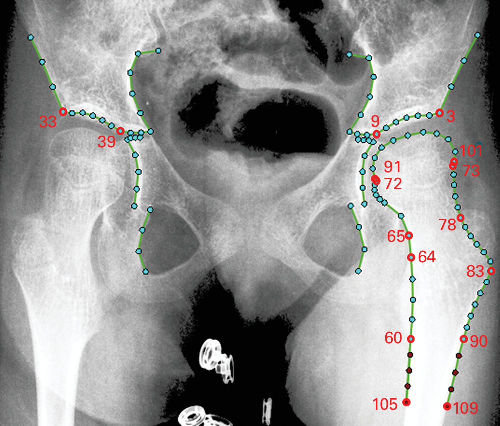
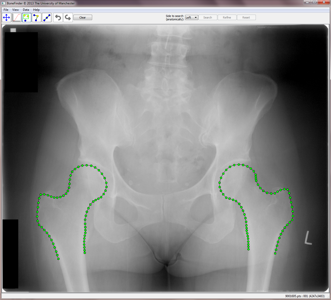
BoneFinder® is a fully automatic software tool to outline and segment skeletal structures from 2D radiographs by placing a set of points along the bone contour or at key landmark positions. It was originally designed for the segmentation of the hip joint but is now also used for other skeletal structures such as the joints of the hand or the knee joint.
BoneFinder® firstly identifies the rough position of the bone in the image and then outlines its contour. To do so, BoneFinder® follows a machine-learning approach. This means that it has been trained on lots of examples in order to learn what to look for in an image, and it now uses this acquired knowledge to identify and segment similar bones in new unseen images. BoneFinder® has been found to lead to very robust and accurate point placements across skeletal application areas.
The resulting point positions can then be used for a range of shape analyses such as building statistical shape models or automatically deriving conventional geometric measurements.
Scientific background: BoneFinder® uses a Hough Forest like approach to detect the structure of interest in the image, and then applies Random Forest Regression-Voting in the Constrained Local Model framework to locally refine all point positions. More details on these methods and relevant references can be found in our peer-reviewed journal publications of international standing in IEEE TMI and IEEE PAMI. The underlying BoneFinder®-technology has been patented: T. Cootes, C. Lindner, M. Ionita. Image processing apparatus and method for fitting a deformable shape model to an image using random forest regression voting. Patent numbers EP 2893491 (approved for GB, FR, DE), US 9928443 (approved for US).
BoneFinder® was written by Claudia Lindner, Tim Cootes and other members of the Centre for Imaging Sciences at The University of Manchester, UK. Funding for the development of BoneFinder® has been received from the Medical Research Council UK, the Engineering and Physical Sciences Research Council UK, Versus Arthritis, the Wellcome Trust and the National Institute for Health Research.
We tested BoneFinder® on a range of different skeletal structures using two-fold cross-validation experiments.
The results show that BoneFinder® achieves state-of-the-art performance across application areas.
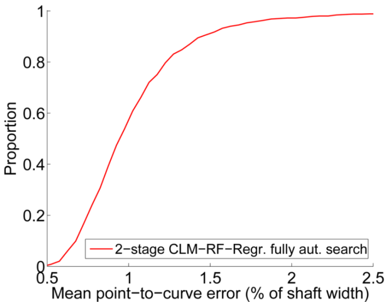
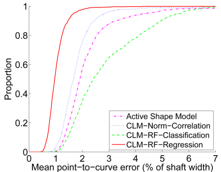
We tested the BoneFinder® hip module on 839 AP pelvic radiographs (527 females, 312 males) using two-fold cross-validation experiments. The aim was to place 65 dense points along the front-view contour of the proximal femur. We report mean point-to-curve errors (where "mean" refers to averaging the errors over all points per image) as a percentage of the shaft width (based on a subset of calibrated images we estimated the latter to be 37mm).
BoneFinder® outperforms alternative matching techniques significantly when starting searching from the mean shape at true pose. It achieves a mean point-to-curve error of less than 0.9mm for 99% of all images (i.e. for 99% of images the error is less than 0.9mm).
BoneFinder® is fully automatic and highly accurate. Without any initialisation it achieves a mean point-to-curve error of less than 0.9mm for 99% of all images. This is equally good as a local search started from the mean shape at true pose (see left plot).
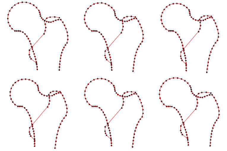
Examples of segmentation results of the fully automatic system (sorted by mean point-to-curve error percentiles): a) median – 0.4mm; b) 91.2% – 0.6mm, highest global search error (i.e. the global search had a success rate of 100%); c) 99.0% – 0.9mm; d) maximal overall error – 2.7mm.
Data shown on this page is copyright (c) 2013 IEEE (DOI: 10.1109/TMI.2013.2258030). Personal use of this material is permitted. However, permission to use this material for any other purposes must be obtained from the IEEE by sending a request to
pubs-permissions@ieee.org.
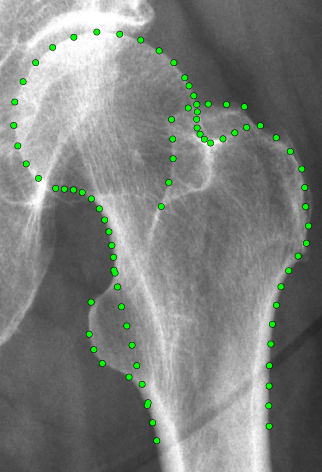
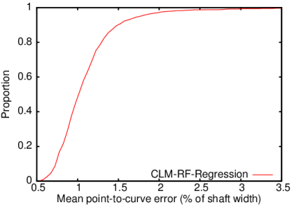
We tested the BoneFinder® hip module on 756 AP pelvic radiographs, aiming to place 81 dense points along the contour of the proximal femur including the trochanters. We report mean point-to-curve errors (where "mean" refers to averaging the errors over all points per image) as a percentage of the shaft width (based on an average of 37mm).
The fully automatic BoneFinder® proximal femur module achieves a mean point-to-curve error of within 1.0mm for 99% of all images (i.e. for 99% of images the error is within 1.0mm).
Segmentation example showing the 95%ile of the fully automatic BoneFinder® proximal femur module with a mean point-to-curve error of 0.7mm for this image. Red points define the femoral shaft width as reference length.
Data shown on this page is copyright (c) 2013 Springer-Verlag (DOI: 10.1007/978-3-642-40763-5_23).
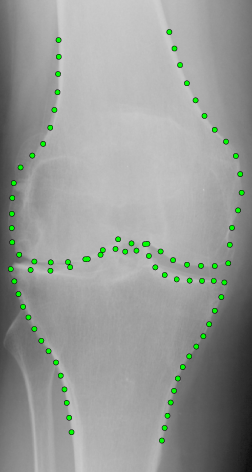
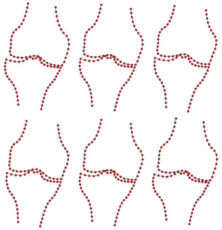
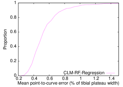
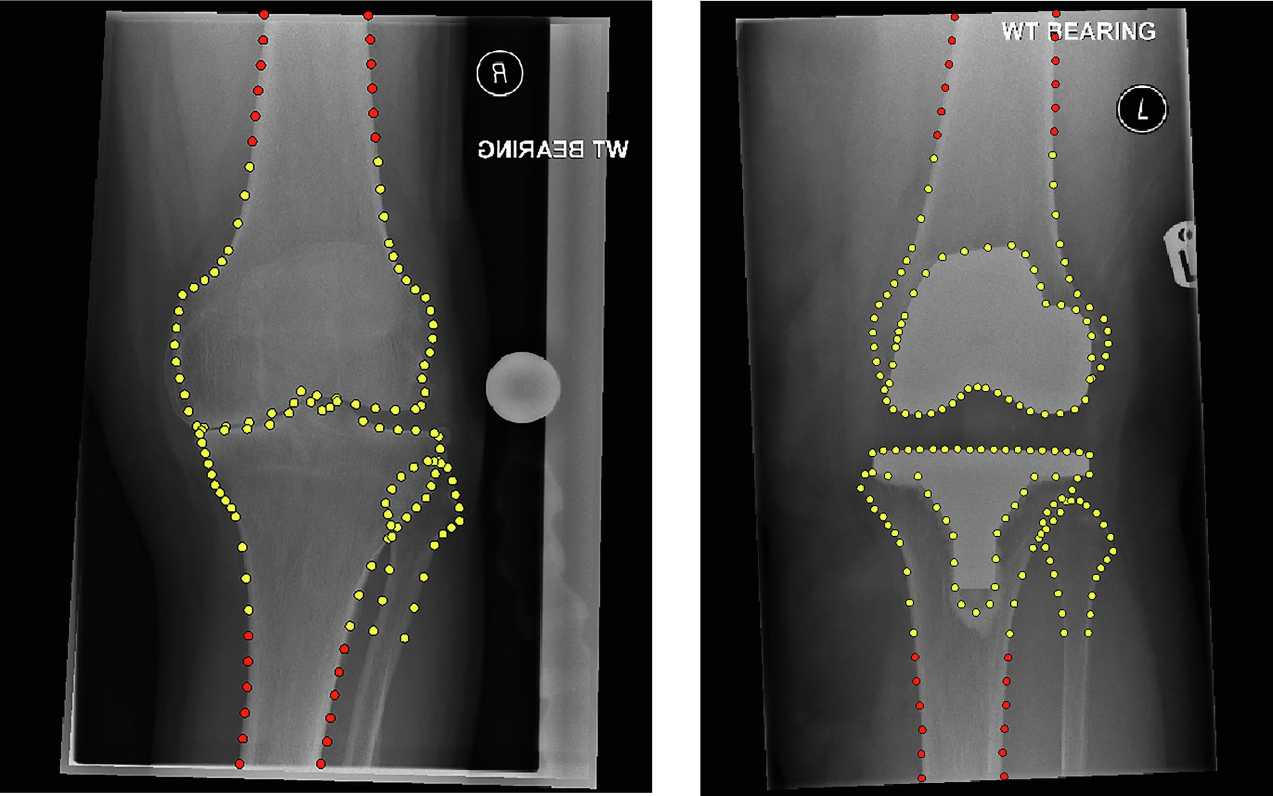
We tested the BoneFinder® knee module on 500 AP knee radiographs, aiming to place 87 dense points along the contours of the distal femur and the proximal tibia. We report mean point-to-curve errors (where "mean" refers to averaging the errors over all points per image) as a percentage of the tibial plateau width (based on an average of 75mm).
The fully automatic BoneFinder® knee module achieves a mean point-to-curve error of less than 1.0mm for 99% of all images (i.e. for 99% of images the error is less than 1.0mm).
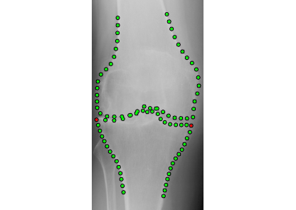
Segmentation example showing the 95%ile of the fully automatic BoneFinder® knee module with a mean point-to-curve error of 0.7mm for this image. Red points define the tibial plateau width as reference length.
Data shown on this page is copyright (c) 2013 Springer-Verlag (DOI: 10.1007/978-3-642-40763-5_23).
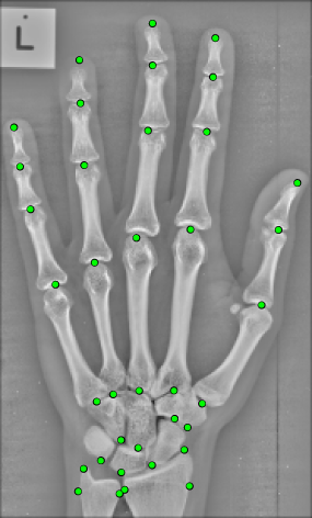
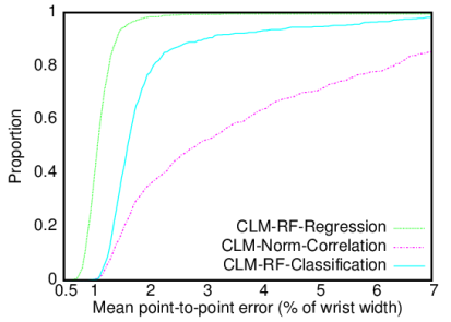
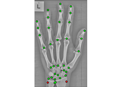
We tested the BoneFinder® hand joint module on 564 hand radiographs, aiming to annotate the joints of the hand (fingers and wrist) with 37 points. We report mean point-to-point errors (where "mean" refers to averaging the errors over all points per image) as a percentage of the wrist width (based on an average of 50mm).
The fully automatic BoneFinder® hand joint module significantly outperforms alternative matching techniques, achieving a mean point-to-point error of within 1.1mm for 99% of all images (i.e. for 99% of images the error is within 1.1mm).
Segmentation example showing the 95%ile of the fully automatic BoneFinder® hand joint module with a mean point-to-point error of 0.8mm for this image. Red points define the wrist width as reference length.
Data shown on this page is copyright (c) 2015 IEEE (DOI: 10.1109/TPAMI.2014.2382106). Personal use of this material is permitted. However, permission to use this material for any other purposes must be obtained from the IEEE by sending a request to
pubs-permissions@ieee.org.
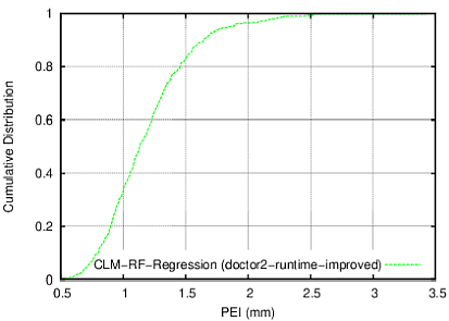
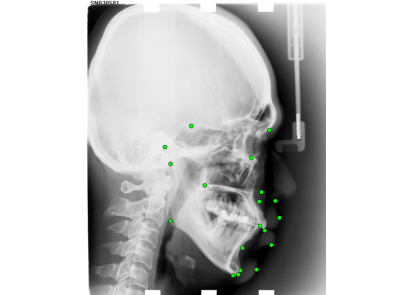
The fully automatic BoneFinder® cephalometric module achieves a mean point-to-point error of within 1.8mm for 95% of all images (i.e. for 95% of images the error is within 1.8mm).
Tracing example showing the 95%ile of the fully automatic BoneFinder® cephalometric module with a mean point-to-point error of 1.8mm for this image.
Data shown on this page is copyright (c) 2019 Springer Nature Limited (DOI: 10.1038/srep33581).
Segmentation example showing the outline of a children's proximal femur. Our BoneFinder® module achieved a point-to-curve-error of less than 4% of the femoral shaft width for 95% of all 1179 images.
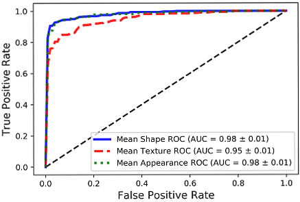
The fully automatic Perthes classification system was able to distinguish between Perthes and healthy childrens' hips with an AUC of 0.98 (SD:±0.01).
Data shown on this page is copyright (c) 2019 Springer Nature Switzerland AG (DOI: 10.1007/978-3-030-11166-3_8).
BoneFinder® is freely available for non-commercial research purposes.
The software can be requested via filling out this licensing form.
Please contact us if you would be interested in the commercial use of the technology.
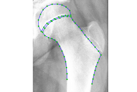
By default, the BoneFinder® research licence comes with the proximal femur model1 as shown on the left. If you are interested in detecting and analysing other skeletal structures please do not hesitate to contact us. We are also working on increasing the number of standard models that will be provided together with the BoneFinder® research licence.
Please cite the following paper when publishing anything resulting from the usage of BoneFinder®:
C. Lindner, S. Thiagarajah, J.M. Wilkinson, The arcOGEN Consortium, G.A. Wallis and T.F. Cootes. "Fully Automatic Segmentation of the Proximal Femur using Random Forest Regression Voting", IEEE Transactions on Medical Imaging, Vol. 32, No. 8, pp. 1462-1472, 2013. DOI: 10.1109/TMI.2013.2258030
1Note that this a front-view proximal femur model that excludes the lesser and greater trochanters and approximates the superior lateral edge from an anterior perspective.
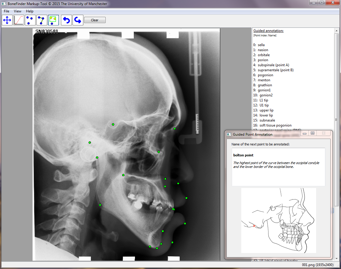
The BoneFinder® Markup-Tool is a convenient tool to manually generate point placements. These could then be used for quantitative shape analyses and/or to train a fully automatic structure-specific BoneFinder® model.
The tool includes guided-annotation which allows the display of the name, description and image of the next point to be placed.
The first version of the BoneFinder® Markup-Tool will be available for download soon. Watch this space.
If you are interested in BoneFinder® and its capabilities please do not hesitate to get in touch by emailing claudia.lindner@manchester.ac.uk
www.manchester.ac.uk/research/claudia.lindner